Some but not all the sub-units have protease activity. These protein compartments contain.
It is the water-based solution in which organelles proteins and other cell structures float.
What are cytosolic proteins. As the most abundant protein in the mitochondrial outer membrane MOM VDAC is known to be responsible for ATPADP exchange and for the fluxes of other metabolites across MOM. It controls them by switching between the open and closed states that are virtually impermeable to ATP and ADP. Tail-anchored TA proteins are membrane proteins that contain an N-terminal domain exposed to the cytosol and a single transmembrane segment near the C-terminus followed by few or no polar residues.
Cytosolic faceof the plasma membraneinclude the cytoskeletal proteins spectrin and actinin erythrocytes Chapter 18 and the enzymeproteinkinaseC. This enzyme shuttles between the cytosoland the cytosolic face of the plasma membrane and plays a role in signal transductionChapter 20. Other peripheral proteins including certain proteins of the.
Besides GAPDH a number of other cytosolic proteins have been found on the surface or to be excreted such as α-enolase Pancholi and Fischetti 1998 glucose-6-phosphate isomerase Hughes et al 2002 glutamine synthetase Suvorov et al 1997 ornithine carbamoyltransferase Hughes et al 2002 fibrinogen-binding protein A of Listeria monocytogenes Dramsi et al 2004 or. Many cytoplasmic proteins can be recruited transiently to cellular membranes. They are collectively known as peripheral proteins.
337 Peripheral proteins may interact with specific membrane lipids via membrane-targeting domains they may contain amphipathic secondary structure or be covalently modified by acyl chains or they may bind to integral membrane proteins. These proteins are degraded by cytosolic proteolytic system present in cell called proteasome. The large 20S proteasome is composed of 14 sub-units arranged in barrel-like structure of symmetrical rings.
Some but not all the sub-units have protease activity. Proteins enter the proteasome through narrow channel at each end. An intracellular protein or protein fraction having a high specific affinity for binding agents known to stimulate cellular activity such as a steroid hormone or cyclic AMP.
Cytosol is the liquid found inside of cells. It is the water-based solution in which organelles proteins and other cell structures float. The cytosol of any cell is a complex solution whose properties allow the functions of life to take place.
Cytosol contains proteins amino acids mRNA ribosomes sugars ions messenger molecules and more. Seperating cytoplasmic and nuclear protein is usually conducted for studing protein distribution especially transcrition factors by western blotting. Tubulin is the most used cytoplasmic marker.
A stainable aggregation of protein lipid or small molecules in the cytoplasm. Moreover protein complexes form in the cytosol allowing the substrate channeling where one product is directly passed to its next step. Some of these complexes also consist of a large isolated central cavity like proteosome.
These protein compartments contain. Protein was determined in both microsomal and cytosolic fractions using the Lowry procedure. Cytoskeletal proteins are recruited from the cytosolic pool to the cell cortex thereby reinforcing it.
Most biotic and abiotic stresses elicit an increase in cytosolic free. Sī tə-sôl -sŏl The fluid component of cytoplasm containing the insoluble suspended cytoplasmic components. In prokaryotes all chemical reactions take place in the cytosol.
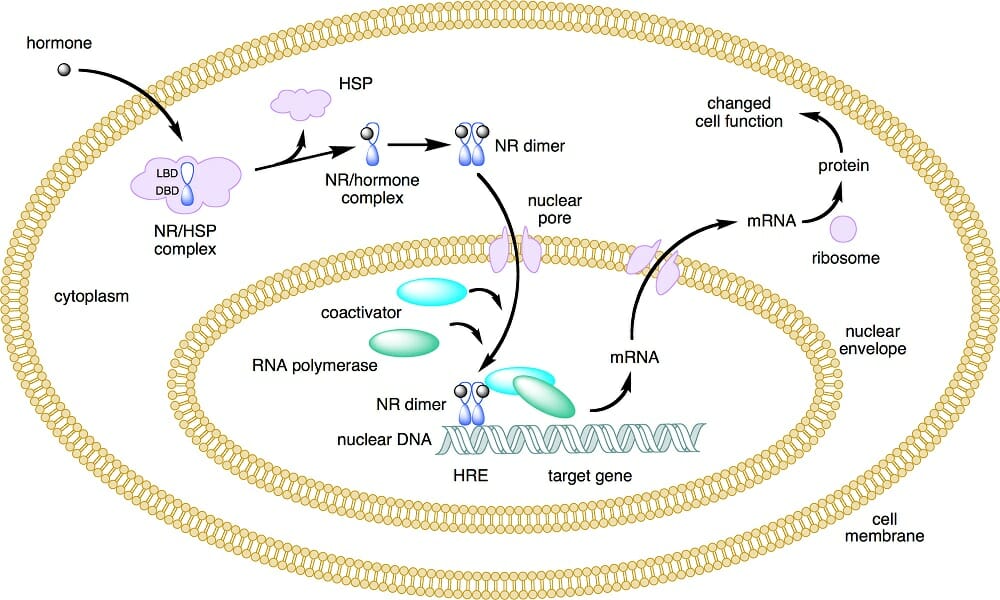
In eukaryotes the cytosol surrounds the organelles. The American Heritage Science Dictionary Copyright 2011. As distinguished from cell surface receptors an intracellular receptor protein especially one for a steroid hormone.
See also nuclear receptor. On the contrary the peripheral proteins on the cytosolic surface are mainly the enzyme protein kinase C which takes part in the signal transduction. In contrast some of the cytosolic peripheral proteins are cytoskeletal proteins.
Similarities Between Transmembrane and Peripheral Proteins.
