According to the morphological characteristic and the similarity of the nucleotide sequence of internal transcribed spacer ITS between r DNAsthe strains producing harpagide or harpagoside were. These slides were labelled and colonies of sporulating species were dried and stored in the.

The slime-molds are morphologically distinct from other fungi in having a body consisting of either cell wall-less amoebae cellular slime molds eg.
Morphological identification of endophytic fungi. Identification of endophytic fungi using traditional techniques Subcultures on different media were examined periodically and identified when isolates sporulated. Slides were mounted in lactophenol and sealed with nail varnish. These slides were labelled and colonies of sporulating species were dried and stored in the.
According to classical mycology most species of endophytic fungi have been described based on their morphological features such as ascospore and peridium morphology gleba colour odour and other organoleptic characteristics. Unique endophytic fungi Debbab et al 2010. Endophytic microorganisms are fungi and bacteria that colonize inter- or intra-cellular spaces of plant tissues during at least one phase of their life cycle as described by Compant and Vacher 2019.
Endophytic microorganisms can protect the hosts against several biotic and abiotic factors such. Among seven plant specimens collected six types of endophytic fungi were morphologically identified as Aspergillus sp. Two different species Penicillium sp Alternaria sp Fusarium sp.
Mangroves morphology Aspergillus sp. The morphological characterization of endophytic fungal isolates was carried out by Tolulope et al 2015 suggested that the endophytic fungal colonies were initially white in color that became. Fifty-two endophytic fungi strains with different colony morphologies were isolated from stems leaves and roots of Huperzia serrata Thunb.
Collected from Bawangling Reserve of Hainan Province in southern China. They were identified mainly based on rDNA ITS sequences and phylogenetic analysis. Isolation and identification of endophytic fungi producing harpagoside and harpagide from Scrophularia ningpoensis.
Article in Chinese. According to the morphological characteristic and the similarity of the nucleotide sequence of internal transcribed spacer ITS between r DNAsthe strains producing harpagide or harpagoside were. The endophytic fungi growing out from the plant tissue were transferred into potato dextrose agar PDA plates and incubate for two to six days.
Continuous plates were subculture until get pure culture. Identification of Endophytic Fungi The isolates of endophytic fungi were identified by the morphology of the fungal culture including colony and medium color Colony characters. The endophytic fungi can be found in any part of the plant such as scale primordial meristem resin ducts petiole buds stem root shoot leaves barks and even in the pneumatophores.
Mangroves act as a host for plenty of endophytic fungal populations. Endophytic fungi in addition to improving plant development may produce antimicrobial substances that can inhibit pathogens. The objective of this study was to isolate and characterize endophytic fungi of Bambusa oldhamii and evaluate in vitro antagonism to Pyricularia oryzaeFungal isolates of B.
Oldhamii shoots were purified and identified by nuclear ribosomal DNA nrDNA. A mangrove endophytic fungus 1403 isolated from the South China Sea Coast which is able to produce griseofulvin and anthracenediones under submerged fermentation was compared with Fusarium genus with the similar morphological characters such as elongated microconidium-producing conidiophores ovoid microconidia and straight to slightly curved macroconidia. The slime-molds are morphologically distinct from other fungi in having a body consisting of either cell wall-less amoebae cellular slime molds eg.
Dictyostelium or a mass of multinucleate protoplasm in which individual cells are indistinguishable acellular slime molds eg. 35 Morphological and molecular identifications of the selected fungus 44 351 Macroscopic identification 44 352 Microscopic identification 45 353 Molecular identification 45 36 Cultivation of fungal isolate and extraction of the crude extract 48 37 Antimicrobial activity tests 49. Thirty isolates of endophytic fungi were isolated from healthy asymptomatic leaves of tea plant Camellia sinensis and identified morphologically based on colony morphology spore shape and size growth and sporulation rate.
Internal transcribed spacer r-DNA sequence analysis supported for molecular identification of all the isolates. Endophytic fungi was isolated from leaf and stem tissue each from Anfas Willard Badam Model Kadari Pancharasi and White sari whereas two fungi were isolated from leaf tissue of KisanBogh and one from stem tissue. The identification of fungi at the genus level was carried out by using macroscopic and microscopic examinations depending on colony color shape hyphae conidia conidiophores and arrangement of.
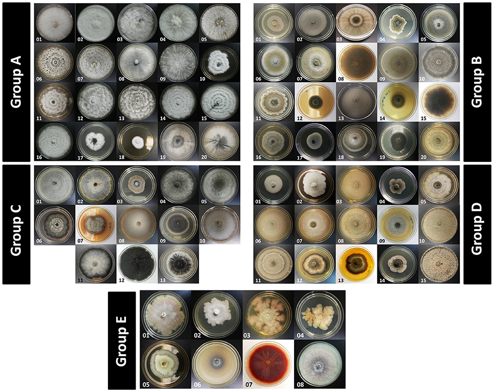
Of endophytic fungi Fig 1. In the present study total fifteen endophytic fungi were isolated from the leaves of four medicinal plants and identified by colony morphology macroscopic and micro-scopic observations. It was found to be Curvularia sp Fusarium sp Penicilluium sp Colletotrichum sp Aspergillus sp Alternaria sp respectively.
Endophytic Fungi Identification. A total of 350 endophytic fungi isolates Supplementary Table S1 were successfully isolated from 300 leaf segments of.