Despite extensive malformations some patients with Chiari II have normal intelligence and can function independently. Note crowding of foramen magnum by the ectopic cerebellar tonsils T and the medulla M.

Click here for Chiari malformation pictures.
Chiari malformation type 1 images. Pictures of Chiari Malformations. Its only natural to want to know what a Chiari malformation looks like. These images will help you understand what a Chiari malformation is and how decompression surgery helps to resolve it.
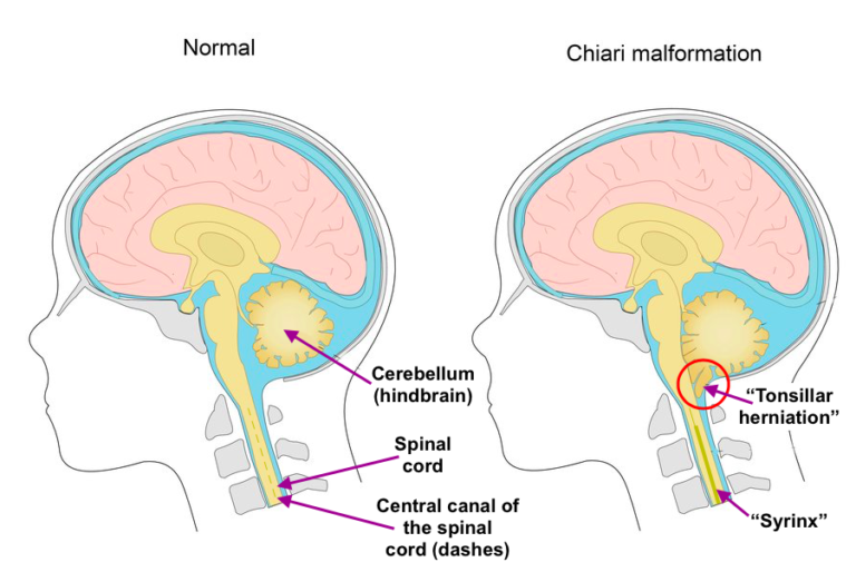
This illustration shows the cerebellar tonsils descending from the skull toward the spinal column creating pressure. Click here for Chiari malformation pictures. You can also find pictures of arnold chiari malformation chiari malformation type 1.
Images of chiari malformation type 1 MRI Chiari I revolutionized diagnostic evaluation for malformation as this method remained previously unrecognized or misdiagnosed as Chiari I could be used to detect malformation. Tonsilposion tonsil configuration and many accompanying abnormalities were shown in sagittal and axial T1 and T2 weighted MRI. 56 righe Chiari malformation type 1 is a structural abnormality of the cerebellum the.
Axial MRI image at the level of foramen magnum in Chiari type I malformation. Note crowding of foramen magnum by the ectopic cerebellar tonsils T and the medulla M. Also note the absence of cerebrospinal fluid.
Life expectancy for Chiari malformation depends on the type. Patients with Chiari type I malformation the mildest form of the condition are typically diagnosed in adulthood and have a normal life expectancy and good outcomes with treatment andor surgery. Despite extensive malformations some patients with Chiari II have normal intelligence and can function independently.
Chiari malformation is considered a congenital condition although acquired forms of the condition have been diagnosed. In the 1890s a German pathologist Professor Hans Chiari first described abnormalities of the brain at the junction of the skull with the spine. He categorized these in order of severity.
See more ideas about chiari malformation chiari awareness. Aug 26 2020 - Explore Amy Leann Dohertys board chiari malformation followed by 1045 people on Pinterest. Chiari malformations are classified by the severity of the disorder and the parts of the brain that protrude into the spinal canal.
Chiari malformation Type I Type 1 happens when the lower part of the cerebellum called the cerebellar tonsils extends into the foramen magnum. Normally only the spinal cord passes through this opening. Chiari Malformation Type 1 Overview Chiari malformations CMs are neurological disorders in which the cerebellum extends out of the skull and into the spinal canal which in turn blocks the flow of cerebrospinal fluid puts pressure on the brainstem and spine and may result in varying degrees of nerve compression.
3299 likes 1 talking about this. Some Chiari memes and a little Chiari poetry. Some are mine some arent.
Clinical findings and magnetic resonance MR images in 68 patients with Chiari I malformations were retrospectively analyzed to identify those radiologic features that correlated best with clinical symptoms. A statistically significant P 03 female predominance of the malformation was observed with a female. Male ratio of approximately 32.