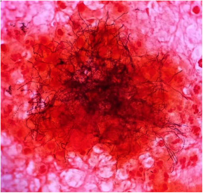
Meyeri which is small and nonbranching all the other species are branching filamentous rods. Schaalia meyeri PIP 2477B is an anaerobe mesophilic gram-positive bacterium that forms circular colonies and was isolated from human purulent pleurisy.

These bacteria are generally anaerobes.
Actinomyces meyeri gram stain. A Gram stain of the specimen is usually more sensitive than culture especially if the patient had received antibiotics. Actinomyces are non-spore-forming Gram-positive rods. Meyeri which is small and nonbranching all the other species are branching filamentous rods.
Although they are gram-positive they often stain irregularly with a beaded appearance non-acid-fast. Slender or slightly curved rods with true branching. Gram stain of the excised sample reveals gram variable filamentous rods.
Therefore it should be distinguished from Nocardia and Streptomyces. On an anaerobic culture Actinomyces meyeri forms creamy-white moist confluent colonies on chocolate agar. However cultures are negative in more than 50 of cases 10.
Actinomyces spp and Propionibacterium propionicus previously Arachnia propionica are members of a large group of pleomorphic Gram-positive bacteria many of which fhave some tendency toward mycelial growth. Both are members of the oral flora of humans or animals. Actinomyces species in particular are major components of dental plaque.
Although actinomycosis is relatively rare at least in Western populations recently reported observations implicating A. Meyeri in brain abscesses and Actinobaculum schaalii currently Actinotignum schaalii in urosepsis and the introduction of advanced microbiological techniques which can identify even very fastidious organisms have resulted in an increased awareness of Actinomyces and other Gram. Actinomyces georgiaeActinomyces gerensceriae and Actinomyces meyerihave been isolated from gingival crevices of periodontally healthy individuals 3 10.
Two new Actinomyces species of oral origin have been described recently. Actinomyces radicidentis from infected root canals 4 and Actinomyces graevenitzii from respiratory tract secretions 19 and infants saliva 21. Actinomycetes in tissue do not stain with the H E stain commonly used for All genera may produce granules.
Actinomyces stain in tissue with Gomori methenamine silver and the Brown and Brenn modification of the Gram stain 8. Most of the literature classifies the tissue response as granulomatous or granulomatoid-like although giant cells and granulomata are rarely seen 39. Sulphur granules are the pathological hallmark of the disease.
Are gram-positive obligate anaerobes known to reside in the mouth and intestinal tract. They are morphologically similar to fungus in that they form filamentous branches. Pathology due to proliferation of organisms usually occurs following injury or trauma to tissue resulting in actinomycosis abscess formation and swelling at the site of infection.
Figure 603 Gram stain of dacryolith showing actinomyces. Actinomyces are mobile or nonmotile chemo-organotrophs requiring preformed organic compounds as a source of carbon and oxidizing organic compounds as a source of energy and can be differentiated by their ability saccharoclastic or inability nonsaccharoclastic to cleave complex sugars. Schaalia meyeri PIP 2477B is an anaerobe mesophilic gram-positive bacterium that forms circular colonies and was isolated from human purulent pleurisy.
Gram stain of the excised sample reveals gram variable filamentous rods. Therefore it should be distinguished from Nocardia and Streptomyces. On an anaerobic cul- ture Actinomyces meyeri forms creamy-white moist confluent colonies on chocolate agar.
However cultures are negative in. Actinomyces from the Greek actinomycete or ray fungus refers to several species of gram-positive non-spore-forming anaerobic bacteria. Human actinomycosis can be caused by any one of 6 species with A.
Israelii named for the author of the first described case in 1891 4 being the most frequently cultured pathogen. Actinomycosis Gram stain Actinomycosis is primarily caused by any of several members of the bacterial genus Actinomyces. These bacteria are generally anaerobes.
In animals they normally live in the small spaces between the teeth and gums causing infection only. Gram stain of the fluid revealed Gram-positive filamentous rods and cultures of the fluid grew Actinomyces species Fig. We analyzed the fluid using a method for clone library sequencing of the 16S ribosomal DNA rDNA gene and Actinomyces meyeri along with other anaerobes Fusobacterium species were detected 10.